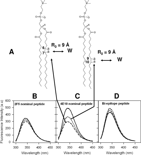
FIG. 6.
Evaluation of membrane insertion of 2F5 and 4E10 nominal epitope and biepitope peptides. (A) Chemical structures of DBr lipids used in the experiment. The positions of DBr labels and Forster distance (R0) requirements for observing 50% quenching efficiency (5) are also shown. (B to D) Tryptophan fluorescence spectra of 2F5 nominal epitope (B), 4E10 nominal epitope (C), and biepitope (D) peptide-liposome conjugates. In each panel, the solid curves show the spectra recorded with no DBr lipids and the dotted and dashed curves show the spectra recorded with 10 mol% 67DBrPC and 910DBrPC liposomes, respectively. a.u., arbitrary unit.

