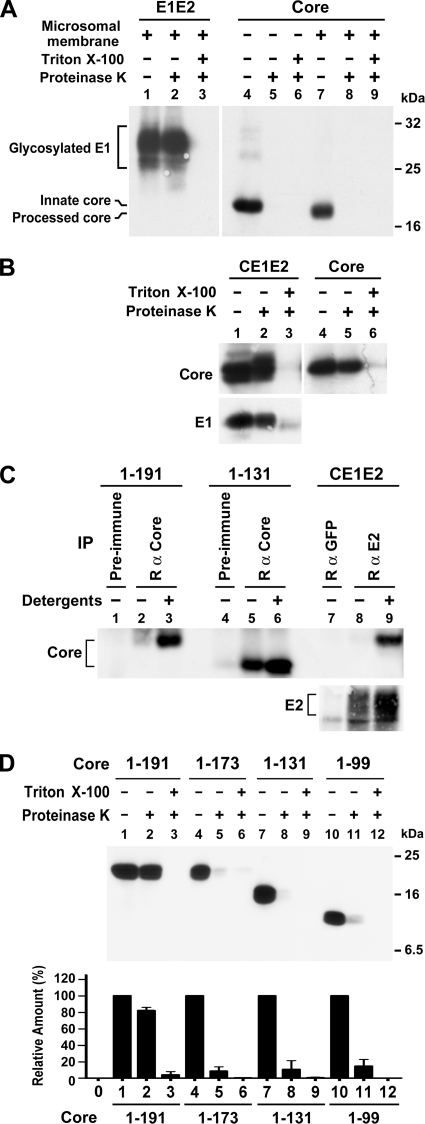
FIG. 2.
Assessment of core envelopment by membrane protection and immunoprecipitation assays. (A) E1E2 and core proteins translated in vitro from pcDNA3-E1E2 and pcDNA3-HACore, respectively, in the presence or absence of canine pancreatic microsomal membranes were subjected to a membrane protection assay. Proteins were resolved by SDS-PAGE, followed by Western blotting with E1- and HA-specific MAbs, respectively. (B) Equal volumes of postnuclear fractions obtained from 293T cells transfected with pCAGGS-HACE1E2 or pCAGGS-HACore were subjected to a membrane protection assay, and samples were analyzed by Western blotting with HA- and E1-specific MAbs, respectively. (C) Postnuclear fractions obtained from 293T cells expressing core or CE1E2 proteins were left untreated or treated with 1% each of NP-40 and sodium deoxycholate prior to immunoprecipitation with the core or E2-specific antibodies as indicated. Incubation with appropriate rabbit preimmune serum or purified rabbit anti-GFP was used as the control. The precipitated antigens were analyzed by Western blotting with MAbs directed against HA and E2, respectively. (D) Postnuclear samples from cells transfected with each core plasmid were analyzed by a membrane protection assay followed by Western blotting with the HA MAb (top panel). The density of bands corresponding to the HA core proteins in each treatment was scanned and quantified. The percentages of core levels detected in samples treated with proteinases K alone or with detergent and protease relative to that of an untreated sample were determined. The diagram represents results from three independent studies with the standard deviation shown (bottom panel).

