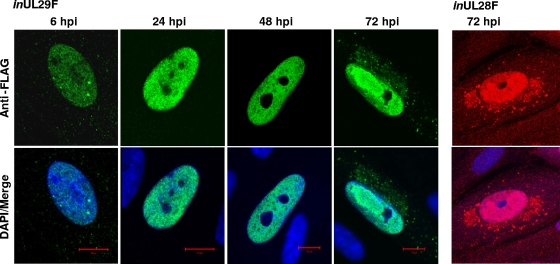
FIG. 3.
Localization of pUL29/28F in fibroblasts during infection. Cells were infected at a multiplicity of 0.5 infectious unit/cell using either inUL29F or inUL28F, fixed at the indicated times, and processed for immunofluorescence using a FLAG-specific antibody (inUL29F, green; inUL28F, red) and DAPI (blue).

