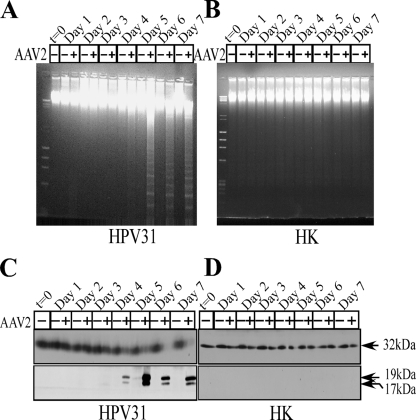
FIG. 1.
AAV2-induced apoptosis in HPV31-infected cells. (A and B) CIN-612 9E (HPV31 positive) (A) and HK (HPV negative) (B) monolayer cultures were synchronized in G1, followed by infection with AAV2 at an MOI of 0.02. Cell pellets were collected each day over a seven-day period. Cells were passaged 1:2 on day 2. DNA laddering assays were performed by isolating low-molecular-weight DNA using standard protocols. Twenty micrograms of DNA was resolved in a 1% agarose-Tris-borate-EDTA gel and stained with ethidium bromide. Detection of caspase-3 cleavage/activation was done by Western blotting. Total protein extracts were prepared and detected as described previously (1). (C and D) Sixty micrograms of total protein extracts from HPV31/AAV2-coinfected cells (C) and HK cells infected with AAV2 (D) were resolved in SDS-polyacrylamide gel electrophoresis (PAGE) gels. To detect the procaspase form, protein samples were resolved in 10% SDS-PAGE gels and detected with caspase-3 rabbit monoclonal antibody (Cell Signaling Technology). To detect the 19-kDa and 17-kDa cleaved caspase-3 forms, protein samples were resolved in 15% SDS-PAGE gels and detected with cleaved caspase-3 rabbit antibody (Cell Signaling Technology). Results shown are representative of three individual experiments. t, time; +, present; −, absent.

