Fig. (3).
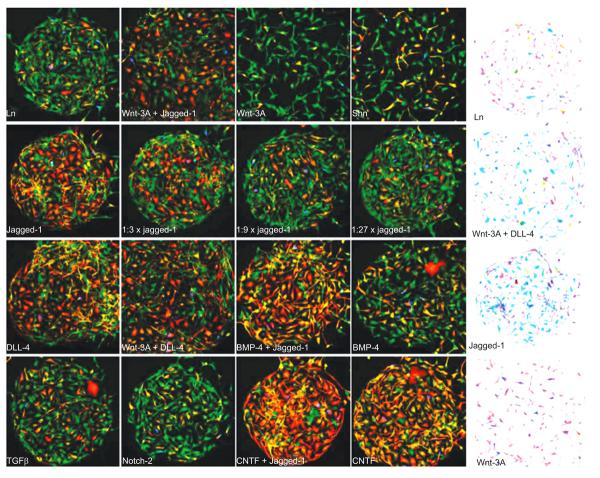
Microenvironment-dependent differentiation and morphology. Human neural precursors were captured and cultured on a printed Ln/ligand array for 70 h under differentiation-promoting conditions. Following the differentiation period, the cells were fixed and counter-stained with GFAP (red), BrdU (blue), TUJ1 (green), and DAPI (not shown). (A) A small portion of the array with 16 different microenvironments each containing a few hundred cells. The balance between TUJ1 and GFAP staining on the reference Ln spot (top left) was skewed toward preferential expression of the neuronal marker TUJ1. This balance was shifted in a spot-dependent manner by some of the signal-containing spots. In particular, spots containing CNTF (bottom right) and Notch ligands (right panels on the 2nd and 3rd rows) led to a dramatic shift toward increased GFAP proportions, suggesting a gliogenic response to Notch stiumulation. Dilution series of Jagged-1 (2nd row panels) revealed dose-dependent response to Notch stimulation. Combination of some gliogenic signals (e.g. Jagged-1 and CNTF) led to further increase in the gliogenic response. A smaller shift toward increased neuronal proportions was observed on Wnt-3A spots. (B) Color inverted images demonstrating spot-dependent morphological differences. Cells that were exposed to a combination of Wnt-3A and a Notch ligand (second spot from the top) exhibited stronger and more elaborated processes compared to Ln alone (top). Typical spot diameter was 400 μm. Fields of view in all panels are identical in size. Wnt-3A-containing spots consistently larger (Figure from reference [37] with permission).

