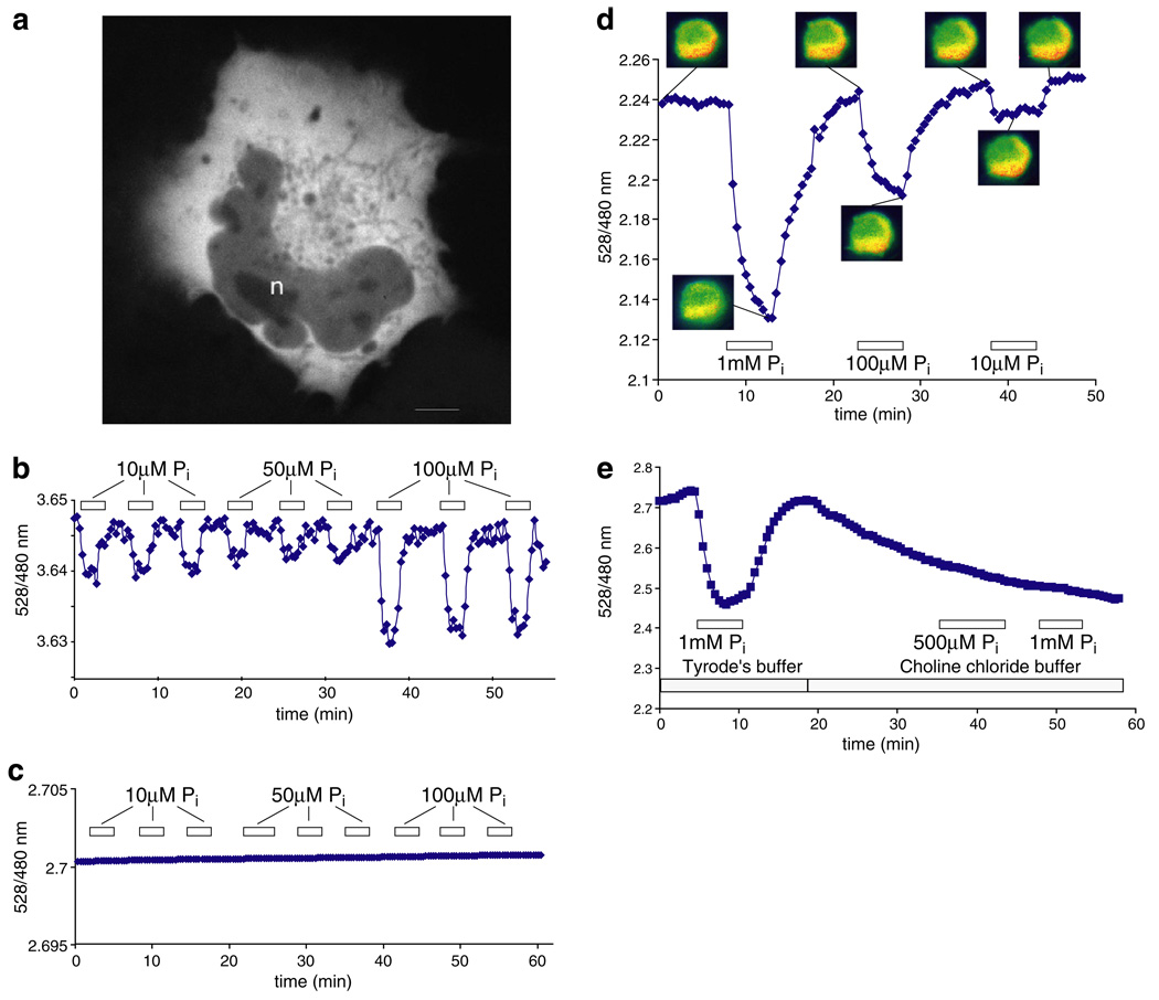
Fig. 4.
FRET ratio change in response to Pi perfusion of Pi-starved CHO cells, and COS-7 cells co-expressing a Na+/Pi cotransporter. (a) Confocal image of a COS-7 cell expressing FLIPPi-30m. Fluorescence is largely excluded from the nucleus (n). The scale bar represents 5 µm. (b) The low-affinity “working” sensor FLIPPi-30m in Pi-starved CHO cells. (c) The high-affinity “control” sensor FLIPPi-5µ is saturated and does not respond to Pi perfusion. (d) The FLIPPi-30m sensor in resting COS-7 cells co-expressing the sodium-dependent phosphate cotransporter PiT2. (e) In sodium-free modified Tyrode’s choline buffer, the response of the sensor to Pi is abolished. The boxed numbers indicate the Pi concentration used for perfusion.

