
Figure 2. The effects of chronic stress in the mPFC are circuit- and estrogen-dependent.
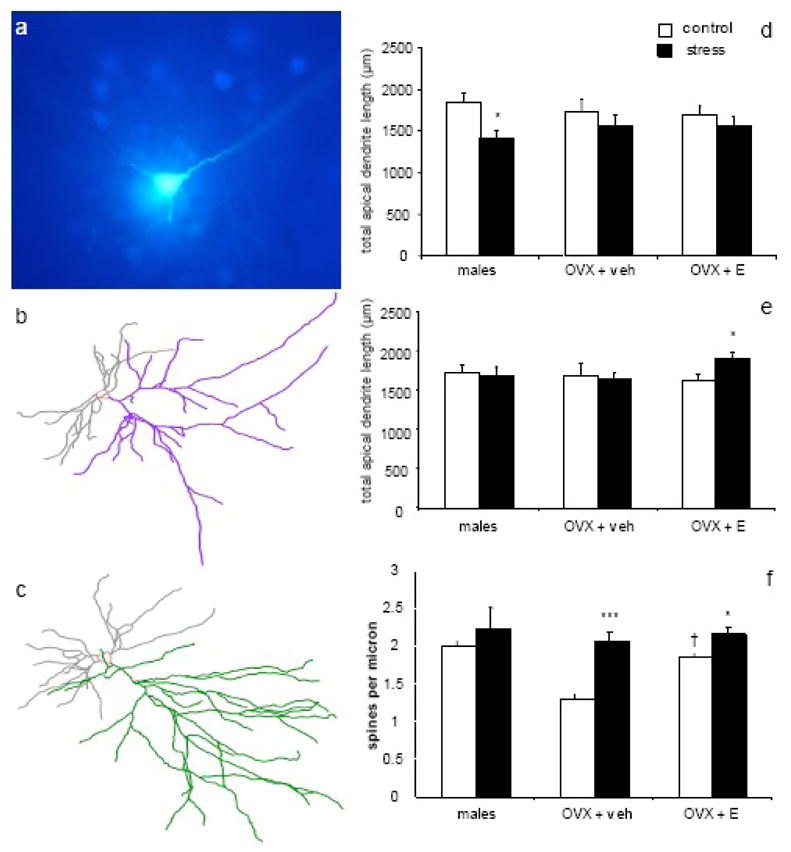
(a) A Lucifer Yellow-filled mPFC pyramidal cell surrounded by FastBlue-labeled cells, demonstrating the technique for selectively loading BLA-projecting neurons. Representative Neurolucida tracings of BLA-projecting neurons from OVX + E control (b) and stress (c) groups. In randomly-selected neurons (d), males, but not females, displayed stress-induced dendritic retraction. However, in BLA-projecting neurons (e), males and OVX + veh showed no stress-induced changes, while OVX + E exhibited stress-induced dendritic expansion. Stress-induced increases in spine density (f) were seen in BLA-projecting neurons of both OVX + veh and OVX + E but not males, and estrogen alone increased spine density in control animals. * p < 0.05 compared to same-group control; *** p < 0.0001 compared to same-group control; † p < 0.05 compared to OVX + veh control. Adapted from (Shansky et al., 2009) and Shansky et al, unpublished observations.