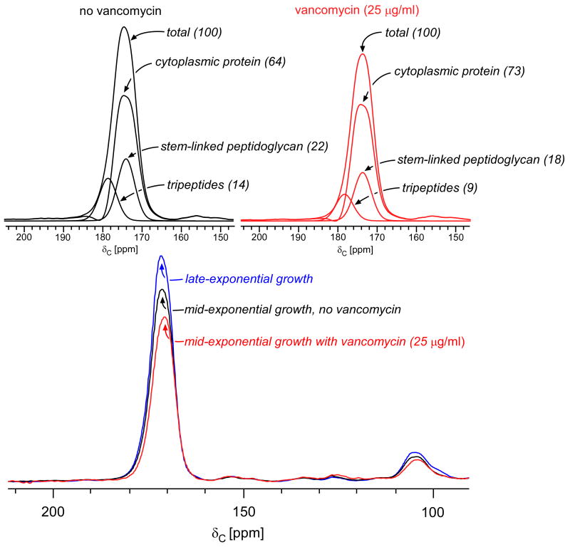
Figure 4.
(Bottom) 13C CPMAS echo spectra of E. faecium whole cells enriched with L-[1-13C]lysine harvested at late-exponential growth (blue), mid-exponential growth (black), or mid-exponential growth in the presence of 25 μg/ml vancomycin (red). The spectra are normalized with respect to the natural-abundance, aliphatic-carbon signal intensities between 0 and 35 ppm. (Top) Deconvolution of the carbonyl-carbon peak for cells in mid-exponential growth, with and without vancomycin. The terminal carboxyl L-Lys in tripeptide peptidoglycan stems has a unique chemical shift of 178 ppm. The remaining peptidoglycan contribution to the peak centered at 175 ppm results from L-Lys stem-linked to D-Ala and was determined by subtracting contributions from cytoplasmic proteins and tripeptides from the total. The cytoplasmic protein concentration is relatively unchanged by drug administration (reference 26). Tripeptides constitute 40% of the peptidoglycan in the untreated control culture (left) and 35% of the peptidoglycan in cells treated with 25 μg/ml of vancomycin (right).

