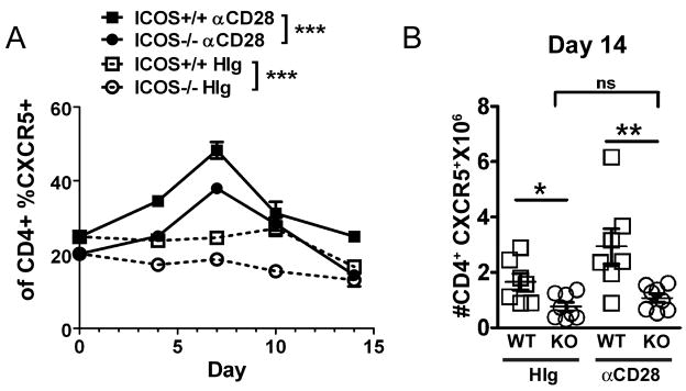
Figure 7. Anti-CD28 cannot sustain an increase in CD4+CXCR5+ T cells (TFH) in the absence of ICOS.
After sensitization with S. mansoni, ICOS+/+ and ICOS−/− mice injected i.p. on day 0 with anti-CD28 or HIg were sacrificed on the days indicated. A, Percentage of spleen CD4+CXCR5+ T cells in αCD28 or HIg treated ICOS+/+ mice or ICOS−/− mice. Significance determined by two way ANOVA as indicated in figure. B, Number of CD4+CXCR5+ T cells found in the spleen on Day 14.

