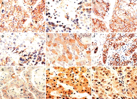
Figure 1.
Immunoreactivity of E-cadherin, α- and β-catenins in HCCs. A and B: Stained for E-cadherin: preserved type (+), reduced type (-); C-E: Stained for α-catenin: preserved type (+), reduced type (-) and staining in the cytoplasm; F-I: Stained for β-catenin: preserved type (+), reduced type (-) and staining in the cytoplasm and the nucleus (× 200).

