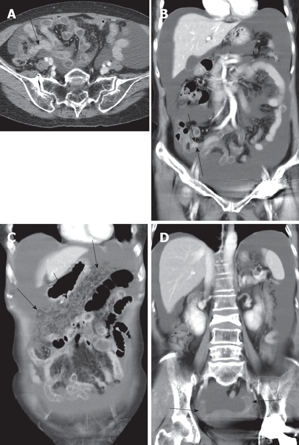
Figure 1.

Contrast enhanced CT axial scan (A) and coronal reformatted image (B) showing massive ascites in abdominal cavity. Appendix is prominently seen with mild thickening (arrowed). Coronal reformatted images (C and D) showing increased reticulonodular densities (arrows in C) along the omentum representing carcinomatosis peritonei with no definite evidence of mass like lesion in adnexa (arrows in D).
