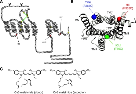
Figure 1.
β2AR single-cysteine constructs and FRET donor–acceptor pair. (A) Three single-reactive cysteines constructs were generated on a minimal cysteine background (Δ5-β2AR). The labelling sites were placed in the first ICL, Δ5-β2AR-T66C, at the cytoplasmic end of the sixth transmembrane segment, Δ5-β2AR-A265C, and helix eight, Δ5-β2AR-R333C. (B) Intracellular 3D view of the distribution of regions chosen for single-cysteine mutants, α-carbons are depicted. (C) FRET donor (λex=549 nm; λem=570 nm) and acceptor pair (λex=650 nm; λem=670 nm).

