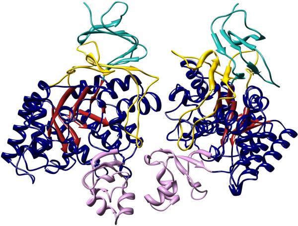
Figure 7.
Homodimeric structure of Bb3285. The (β/α)8-barrel is colored with red β-strands and dark blue α-helices and adjoining loops. The first insertion domain (from residue 287 to 344) containing the substrate specificity loop is colored pink. The second insertion domain consisting of residues 5−61 and 413−478 are colored cyan and yellow, respectively. Molecular graphics images in Figures 7 and 9 were produced using the UCSF Chimera package from the Resource for Biocomputing, Visualization, and Informatics at the University of California, San Francisco, supported by NIH P41 RR-01081 (38).

