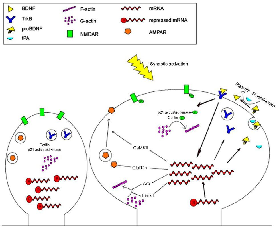
Figure 2. Mechanisms of BDNF induced synaptic plasticity.
In the non-stimulated state, dendritic spines remain “silent”: mRNAs are repressed in RNA granules, TrkB and AMPA receptors are held intracellularly, and actin dynamics leave the spine head in an immature form. Following synaptic activation, repressed mRNAs are disinhibited, TrkB is inserted into the plasma membrane, pro-BDNF and tPA are packaged either together or separately and released into the synaptic cleft, pro-BDNF is converted into BDNF by plasmin, and BDNF binds to TrkB on the local dendritic membrane. Activation of TrkB by BDNF increases translation of CaMKII, GluR1, Arc, and Limk1, leading to increased the formation and membrane insertion of the AMPA receptor and increased actin polymerization. TrkB signaling also induces phosphorylation of NMDA receptors, synapsin-1, p21 activated kinase, and cofilin, thus increasing receptor activity, vesicle-plasma membrane fusion and neurotransmitter release, and polymerization of actin, respectively.

