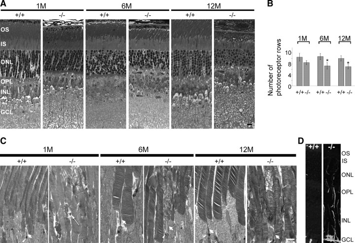
Figure 3.
Photoreceptor abnormalities in Rp1L1−/− retinas. A, Light micrographs of epoxy-embedded sections of central retinas of Rp1L1 mutant mice at ages 1 (1M), 6 (6M), and 12 (12M) months. Scale bar, 5 μm. The OS and ONL are significantly thinner in retinas of 6-month-old Rp1L1−/− mice than in retinas of wild-type littermates. B, Average numbers of photoreceptor rows in the ONL of central retinas located 300–400 μm from the optic nerve at ages 1, 6, and 12 months as described before (Gao et al., 2002). Group mean values and SDs (bars) are shown. Progressive degeneration of photoreceptors (manifested as the numbers of photoreceptor rows) in Rp1L1−/− mice is much milder than in Rp1−/− mice (Gao et al., 2002). C, Ultrastructural (transmission electron) micrographs of OSs at ages 1, 6, and 12 months. Scale bar, 2 μm. Note the appearance of vacuoles in several OSs (1M) and abnormal discs in isolated OSs flanked by normal-appearing OSs (1, 6, and 12M). Interestingly, the abnormal discs that are in the middle or basal segment of the OS and are bordered by normal-appearing discs in the same OS (6 and 12M). D, GFAP induction in retinal Müller glia in Rp1L1−/− retinas at 3 months (3M). Rp1L1+/+ (left) and Rp1L1−/− (right) retinas were stained with monoclonal anti-GFAP antibodies and fluorescein-conjugated secondary antibody. Scale bar, 10 μm.

