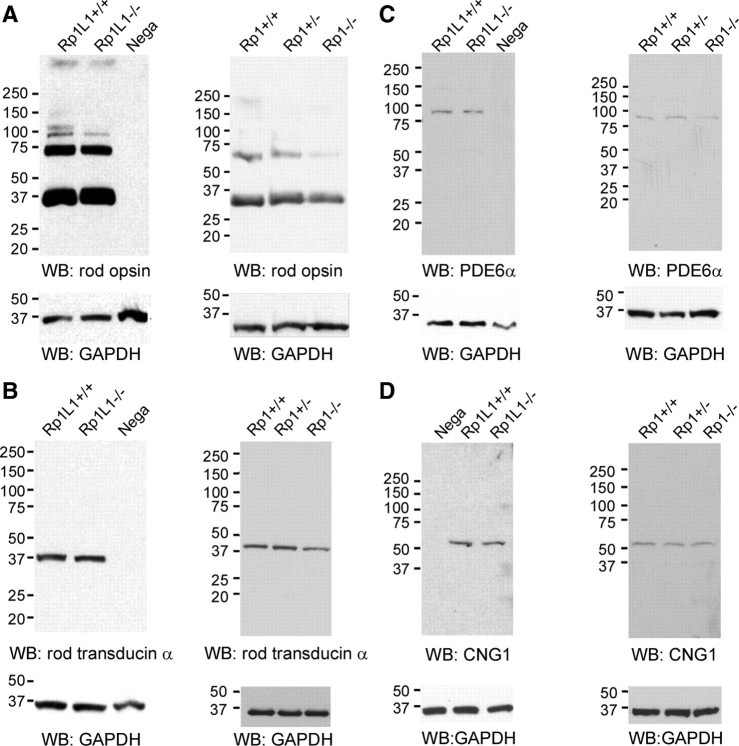
Figure 8.
Expression of other phototransduction proteins, rod opsin (A), rod transducin α (B), PDE6α (C), and CNG1 channel (D), in Rp1+/−, Rp1−/−, Rp1L1−/−, and wild-type mice that had been exposed to ambient light for ∼8 h. The retinas from Rp1L1−/− and wild-type mice at P46–P47 were analyzed by SDS electrophoresis and probed with antibodies as shown (left). Cerebellum whole-cell lysates were used as a negative control. Because of the severe degeneration of photoreceptors, the whole-retina lysates from Rp1+/−, Rp1−/−, and wild-type mice were analyzed at P21 (right). An anti-GAPDH antibody was used for the detection of the loading control. Note slight reductions of rod opsin, rod transducin α, and PDE6α protein in Rp1−/− retinal lysates, attributable to the shortened OS in Rp1−/− rods, as described by Liu et al. (2004). WB, Western blot.

