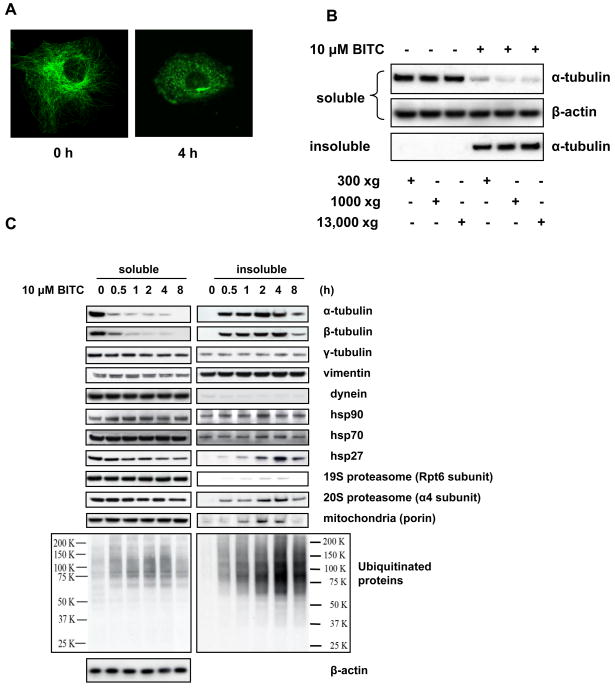
Figure 1. ITCs induce formation of aggresome-like structure in cells.
A, BITC treatment disrupts microtubule network and induces tubulin aggregates. Immunofluorescent staining images of α-tubulin before and after HeLa cells were treated with 5 μM BITC for 4 h. B, ITC-induced tubulin-containing complex can be spun down at low centrifuge speed. HeLa cells were treated with 10 μM BITC for 2 h. The non-ionic detergent soluble and insoluble fractions were separated at various centrifugation speeds and immunoblotted separately for α-tubulin. C, Some aggresome marker proteins are enriched in the ITC-treated tubulin aggregates. HeLa cells were treated with 10 μM BITC for up to 8 h. The soluble and insoluble fractions of the cell lysate was immunoblotted for both α- and β-tubulin and aggresome markers γ-tubulin, vimentin, dynein, HSP90, HSP70, HSP27, proteasome components, mitochondria marker, and ubiquitinated proteins.

