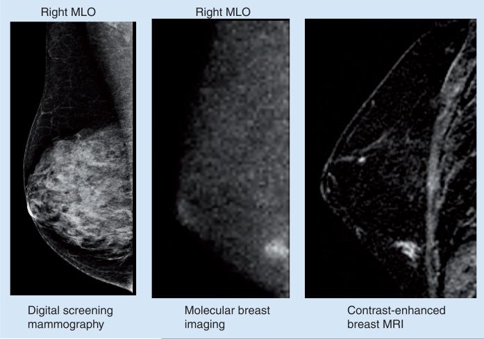
Figure 3. Right MLO views obtained by digital mammography, molecular breast imaging and contrast-enhanced MRI.
Screening mammogram was interpreted as negative for disease. Molecular breast imaging indicated a small lesion in the lower quadrant of the right breast. Lesion was also seen on contrast enhanced breast MRI and was confirmed at surgery as a 9 mm ductal carcinoma in situ.
MLO: Mediolateral oblique.

