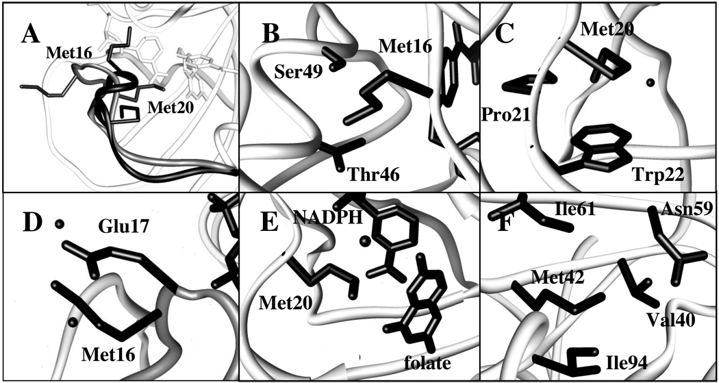
Figure 4.
Environment of Met residues in crystal structures of DHFR bound to folate (PDB 1rx7) or to and folate and NADP+ (PDB 1rx2). (A) Superposition of Met20 loop in the occluded (black) and closed (grey) states with side chains of Met16 and Met20 shown; (B) Met16 in occluded state (His45 backbone carbonyl not shown for clarity); (C) Met20 in occluded state; (D) Met16 in closed state; (E) Met20 in closed state (the folate (p-aminobenzoyl)glutamate group has been omitted for clarity); (F) Met42 (same environment in occluded and closed states). Panels (B) – (F) include crystallographically observable water molecules that are within 4 Å of the labeled ε-methyl group.

