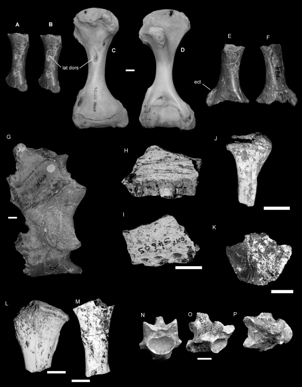
Figure 3. Varanus komodoensis (Neogene, Australia).
A–B, E–G. Pliocene V. komodoensis (Australia)A–B. QMF 53955, partial left humerus in dorsal view showing position of insertion for the latissimus dorsi (lat dors). C–D. Left and right humerus of a modern V. komodoensis (NNM 17504). E–F. QMF 53954, partial right humerus in ventral and dorsal views, showing the position of the ectepicondyle (ect). G. QMF 866, partial scapulocoracoid. H–P. Pleistocene V. komodoensis (Australia). H–I. QMF 54605, partial left maxilla in lingual and labial views. J. QMF 54606, partial right quadrate in anterior view. K. QMF 54607, supraoccipital bone in posterior view. L. QMF 54608, proximal left tibia. M. QMF 54604, ulna diaphysis. N–P. QMF 1418, proximal mid-caudal in cranial, oblique lateral and dorsal views. Scale bar = 1 cm.

