
Figure 2.
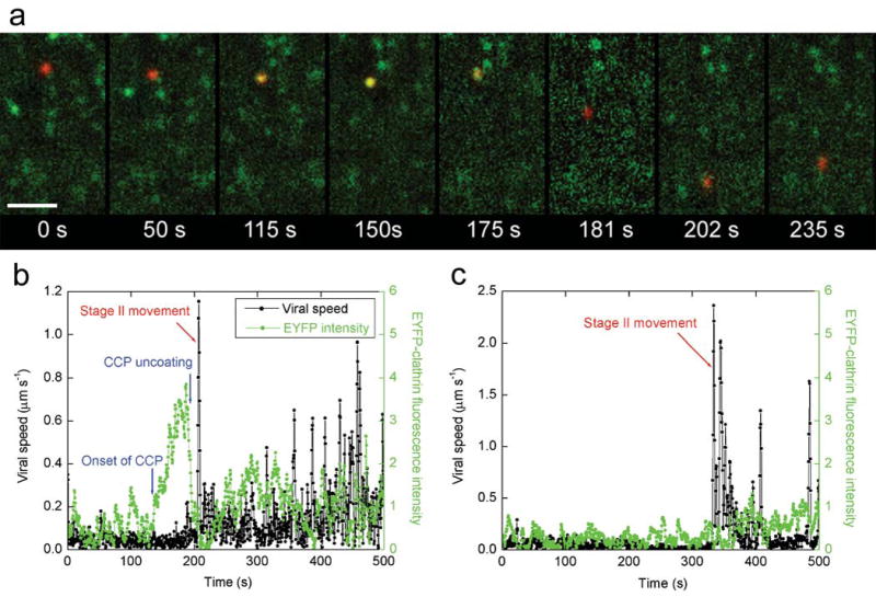
Internalization of influenza viruses via multiple pathways. (a) Snapshots of a virus internalized via a CCP. A live movie of this virus is available as Supplementary Video 3. Scale bar: 10 μm. t = 0 s: the virus (red) binds to the cell. t = 50 s: the virus is undergoing stage I movement. t = 115 s: a CCP labeled with EYFP (green) begins to form at the virus site. t = 150 s, the clathrin coat reaches its peak fluorescence intensity. t = 175 s: the clathrin coat rapidly disassembles. t = 181 seconds: the virus is transported towards the nucleus on a microtubule (stage II movement). t = 202 s: the virus enters stage III transport involving both plus- and minus-ended-directed motilities on microtubules. t = 235 s: the virus continues stage III movement. (b) The time-trajectories of a virus internalized via de novo formation of a CCP. Black symbols are the velocity time-trajectories of the virus. Stage II movement is identified as the rapid unidirectional translocation from the cell periphery to the perinuclear region (red arrows). Green symbols are the integrated fluorescence intensity of EYFP-clathrin associated with the virus. (c) The time-trajectories of a virus internalized without association with a clathrin-coated structure. Symbols are as defined in b. Live movies of the two viruses in b and c are also available (Supplementary Videos 4 and 5).