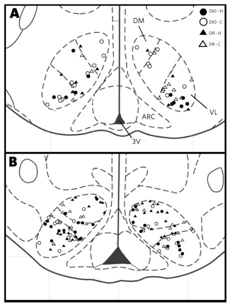
Figure 2.
Drawing of the mediobasal hypothalamus summarizing the location of all VMH neurons in this study, including neurons from both the DIO (open symbols) and DR (filled symbols) animals. The drawings are based on Paxinos and Watson [49]; Panel A is bregma −2.30 and Panel B is bregma −3.80. Abbreviations: f, fornix; 3V, third ventricle.

