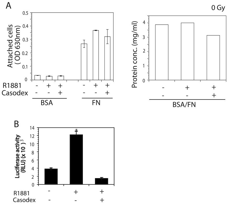
Fig. 1.
Effect of androgen stimulation on LNCaP cell adhesion to FN. LNCaP cells were cultured in 2% CS-FBS containing culture medium for 24 hrs followed by stimulation with 1 nM R1881 for another 24 hrs. Bicalutamide (5 μM) were added into 2% CS-FBS contained culture medium 1 hr before addition of R1881. For adhesion assay, cells were detached and plated onto wells coated with 10 μg/ml FN or BSA (as a control). After 3 hrs of incubation at 37°C, cells were stained with crystal violet. Measuring absorbance at 630 nm scored cell attachment. The same volume of cell suspensions was collected within an experiment and cells were lysed. Protein concentrations of cell lysate were used as a loading control. A: No change in adhesion was observed when LNCaP were stimulated with ethanol or R1881 in the presence or absence of Bicalutamide (left panel). Protein concentrations from the same amount of cells from each treatment for adhesion assay were determined using BSA assay to show the equal cell loading in adhesion assay (right panel). B: LNCaP cells were transiently transfected with plasmid containing ARE luciferase constructs and β-gal. Cells were treated with 1 nM R1881 only or with R1881 in the presence of Bicalutamide (5 μM). AR activity in LNCaP cells was determined by luciferase activity.

