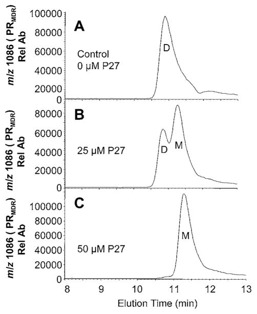
Figure 4. Effect of P27 on PRMDR elution profile and PR activity.
PRMDR (1 μM) was incubated for 16 h at 37 °C in 150 mM ammonium acetate buffer containing 100 μg · ml−1 BSA. Samples (8 μl) were separated by size-exclusion chromatography and elution was monitored by MS for the PRMDR-specific ion (m/z 1086, 10+). (A) Treatment with 0 μM P27 (elution time 10.7 min), (B) 25 μM P27 (elution times 10.8 and 11.3 min) or (C) 50 μM P27 (elution time 11.3 min). The dimeric (D) and monomeric (M) forms of PRMDR are indicated in the Figures. PR activity of each sample was also measured following the incubation. PR activity was (A) 2.6 RFU min−1 · μg−1 (where RFU is relative fluorescence units) (B) 1.85 RFU min−1 · μg−1 and (C) 0.0 RFU min−1 · μg−1 PRMDR. Rel Ab, relative ion abundance.

