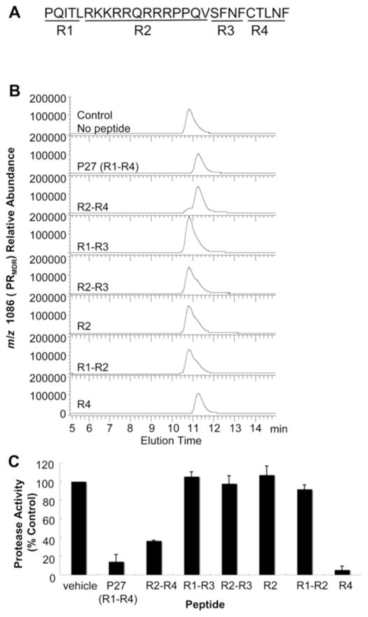
Figure 6. Effect of P27 and P27-related peptides on PRMDR elution profile.
PRMDR was incubated at 1 μM for 16 h at 37 °C with 50 μM of each peptide. Samples were then analysed by size-exclusion chromatography. (A) P27 sequence with the four major peptide regions (R1–R4) indicated. (B) The elution profile for PRMDR by MS of the PRMDR-specific ion (m/z 1086, 10+) is shown for each sample The total area corresponding to PRMDR was: no peptide, 6.1 × 10⁶; R1–R4, 1.6 × 10⁶; R2–R4, 2.9 × 10⁶; R1–R3, 6.5 × 10⁶; R2–R3, 6.9 × 10⁶; R2, 7.1 × 10⁶; R1–R2, 7.7 × 10⁶; and R4, 3.9 × 10⁶. Peak elution times were 10.8 min for control, R1–R3, R2–R3, R2 and R1–R2, and 11.3 min for R1–R4, R2–R4 and R4. (C) Corresponding PR activity for each sample following peptide treatment. Results are means ± S.D. (n = 3).

