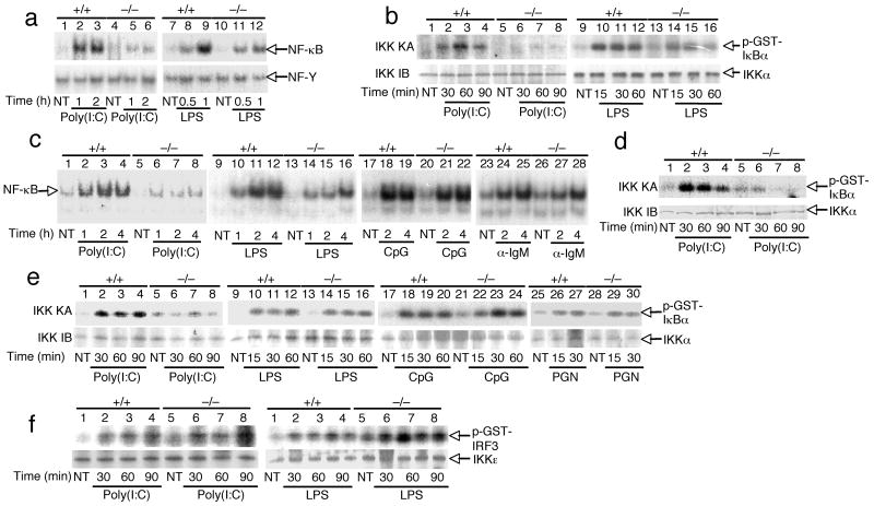
Figure 4. Peli1 is essential for IKK-NF-κB activation by TLR3.
(a) Primary Peli1+/+ and Peli1−/− MEFs were unstimulated (NT) or stimulated with poly(I:C) (50 μg/ml) or LPS (1 μg/ml). Nuclear extracts were subjected to EMSA for detection of NF-κB and NF-Y. (b) MEFs were treated as indicated and subjected to kinase assay (KA) by isolating IKK holoenzyme by IP using anti-IKKγ and using GST-IκBα(1-54) as substrate (upper). Kinase assay membrane was subjected to IB using anti-IKKα (lower). (c,d) Peli1+/+ and Peli1−/− B cells were stimulated as indicated and subjected to EMSA (c) and kinase assays (d). (e) Peli1+/+ and Peli1−/− splenocytes were stimulated as indicated and subjected to kinase assays as described in b. (f) Primary Peli1+/+ and Peli1−/− MEFs were stimulated as in A. IKKε was isolated by IP and subjected to kinase assays using GST-IRF3 as substrate. Data are representative of 3 or more independent experiments.

