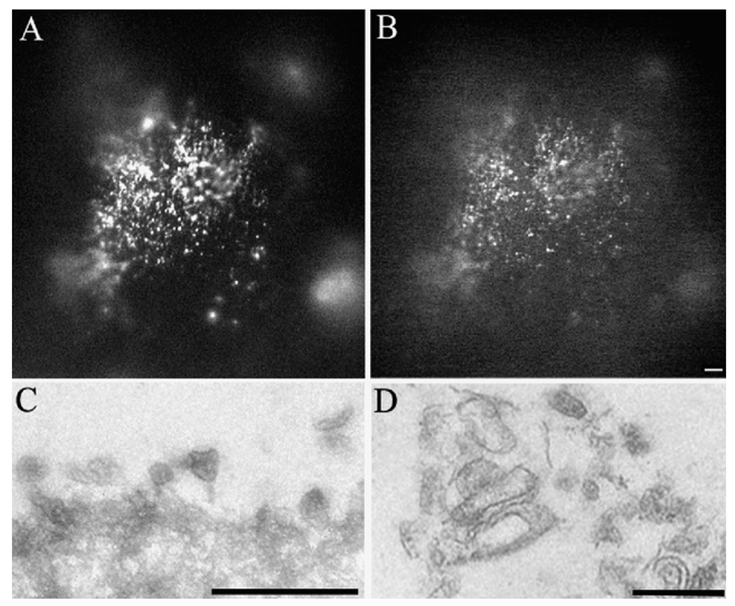
Figure 72.1. Morphological analysis of isolated WGA-beads with mouse RPE microvilli on their surface.
WGA beads scraped off the mouse eyecups were reacted with antibodies to protein kinase A regulatory subunit alpha II (PKARII) (A) and protein kinase A regulatory subunit I (PKARI) (B). Alternatively, the isolated beads were fixed in 2.5% glutaraldehyde, and processed for transmission electron microscopy (TEM). Low (C) and high (D) magnification of these beads revealed extensive surface areas covered by the RPE microvilli. Bars = 100µm (A, B), 1µm (C) and 0.5µm (D).

