
Figure 2. Inflamed Giα2-/- crypts are hypoxic.
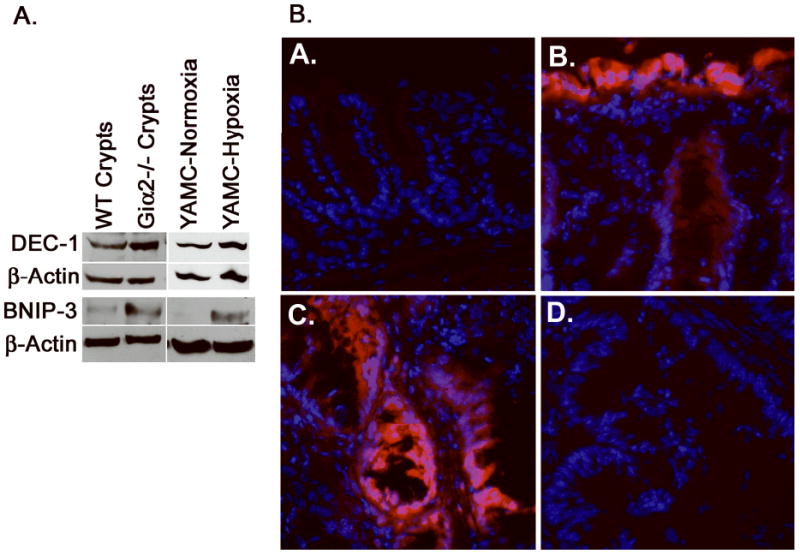
(A), YAMC cells grown in 1% O2 (right) upregulate the hypoxia markers DEC-1 and BNIP3, which are also upregulated in purified crypt lysates from colitic Giα2-/- mice (left). The data shown are representative of three separate YAMC and crypt isolation experiments. (B), Frozen sections from EF5-treated WT and Giα2-/- mice (age 20-27 weeks) were stained with Cy3-conjugated anti EF5. WT colon (A) had minimal EF5 staining, while colitic Giα2-/- mucosa (B) was profoundly hypoxic in surface epithelium and in some deeper crypt cells. EF5 staining of Giα2-/- cancers (C.) was patchy, and involved primarily malignant epithelial cells and the adjacent stroma. As a negative control, pre-adsorbed anti EF-5 (D.) revealed no staining.