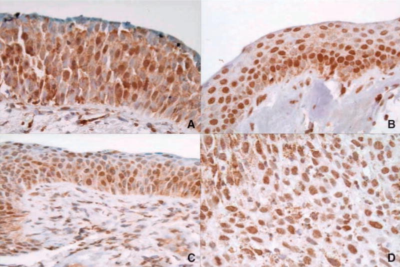
Figure 1.
VDR expression across various lung histologies. A. Normal. B. Metaplasia. C. Dysplasia. D. Squamous cell carcinoma. In many samples, VDR was expressed in a large percentage of the cells, in multiple layers of the bronchial epithelium. Cytoplasmic expression decreased with increasing histologic grade.

