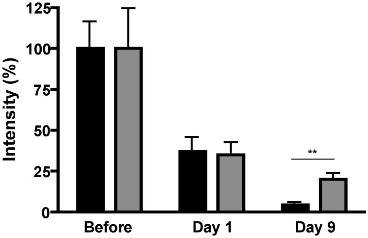
Figure 2.
Prosaposin protein levels of the nerve proximal to the transection site in control (black) and diabetic (grey) rats before and 1 or 9 days after nerve transection. Each lane was initially normalized to sample actin levels and then to the mean intensity of control nerve segments collected during transection (labeled before) that were present on the same blot to allow for inter-blot comparisons. Data are mean ± SEM (n = 8 to 10 per group). **, p < 0.01 by unpaired t-test.

