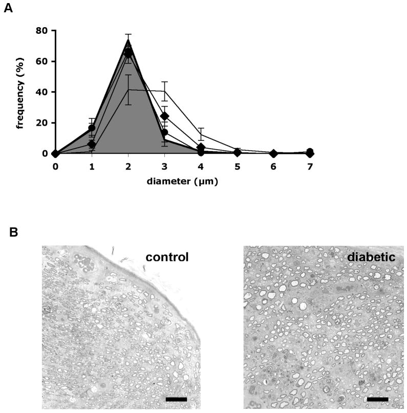
Figure 5.
(A) Axonal size: frequency distribution in the sciatic nerve distal to the crush site measured 1 month after the injury in control rats (grey area under curve), control rats treated with prosaptide TX14(A) following crush injury (filled circles), diabetic rats (line without markers) and diabetic rats treated with prosaptide TX14(A) following crush injury (filled diamonds). Data are group mean ± SEM for each size bin. (B) Light microscopic images illustrating nerve distal to the crush site from control and diabetic rats used for morphometric analysis. Bar = 60 μm.

