Figure 1.
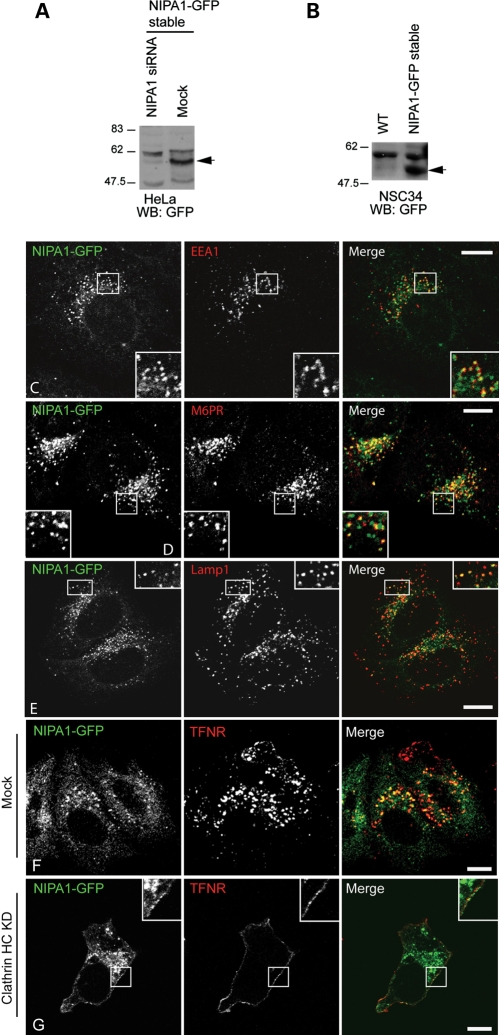
NIPA1-GFP localizes to endosomes. (A) Immunoblot showing expression of a band (arrow) of approximately the size predicted for NIPA1-GFP (∼61 kDa) in the NIPA1-GFP HeLa stable cell line, but not in similar cells transfected with pooled siRNA against NIPA1. (B) Immunoblot showing expression of a band of approximately the size predicted for NIPA1-GFP (arrow) in the NIPA1-GFP NSC34 stable cell line, but not in wild-type (WT) NSC34 cells. (C–E) Stably expressed NIPA1-GFP co-localizes in HeLa cells with early endosome antigen (EEA1; early endosomes; C), mannose 6-phosphate receptor (M6PR; late endosomes; D) and Lamp1 (lysosomes; E). Although results are shown for a mixed stable cell line, similar findings were obtained with individual clonal cell lines (data not shown). (F and G) Clathrin heavy chain (HC) knock-down redistributed NIPA1-GFP to the plasma membrane. (F) Control cells stably expressing NIPA1-GFP in which mock siRNA transfections were carried out. Minimal NIPA1-GFP or transferrin receptor (TFNR) is present at the plasma membrane. However, when clathrin HC is depleted by siRNA knock-down (G), TFNR was redistributed, as expected, to the plasma membrane. NIPA1-GFP was also partially redistributed to the plasma membrane (see inset higher magnification box). In these and subsequent micrographs, the right-hand panels show the merged images; the colour of each marker in the merged image is shown by the colour of its lettering in the non-merged panels. Scale bars: 10 µm in these and subsequent micrographs.

