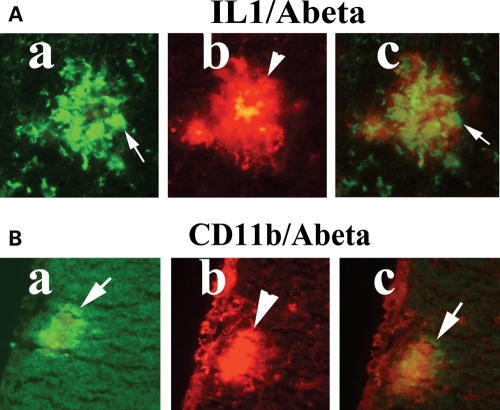
Figure 3.
Double-labeling immunofluorescence analyses of Aβ deposit and microglia marked by IL1β (A) and CD11b (B) in representative 10-month-old Tg2576 mouse. Aβ deposits are associated with microglia. (Aa) Immunoreactivity of IL1, (Ab) surrounded by Aβ deposit in the same section and (Ac) overlay of both IL1 and Aβ deposit. Arrows indicate immunoreactivity of IL1 (in white), and the arrowhead indicates Aβ deposit. (Ba) Immunoreactivity of CD11b, (Bb) surrounded by Aβ deposit and (Bc) overlay of both CD11b and Aβ deposit. Arrows indicate immunoreactivity of CD11b (in white), and the arrowhead indicate Aβ deposit.

