Figure 2.
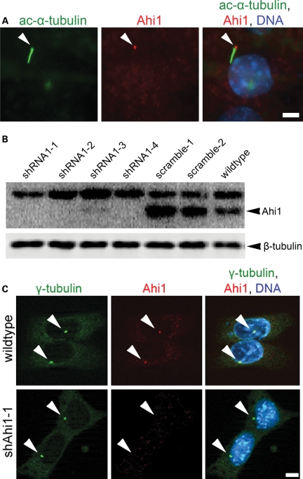
Ahi1 localizes to the basal body of the primary non-motile cilium and its expression is lowered in Ahi1-knockdown IMCD3 cells. (A) Serum-deprived IMCD3 cells have robust cilium formation (one/cell). Ciliated IMCD3 cells were stained with acetylated α-tubulin (green), a ciliary axoneme marker, and for Ahi1 (red). Arrowheads indicate the localization of Ahi1 to the base of a primary cilium. DNA is visualized with Hoechst 33258 (blue). For evaluation of knockdown efficiency of Ahi1, IMCD3 cells stably expressing shRNAi against Ahi1 were established and analysed for Ahi1 protein expression by (B) western blotting and (C) immunostaining. (B) Protein levels of Ahi1 in four independent Ahi1-knockdown IMCD3 stable clones, two control shRNAi scramble stable clones and wild-type cells were analysed by western blotting. Ahi1 (130 kDa, lower band) can be detected in scramble control and wild-type cells, but not in cells expressing a shRNAi against Ahi1. The top band is considered to be a non-specific band (34). (C) Immunostaining for γ-tubulin (green), for Ahi1 (red) and for DNA (Hoechst 33258 (blue)) was performed in wild-type IMCD3 cells and in Ahi1-knockdown cell line 1 (shAhi1-1). Scale bars = 5 µm.

