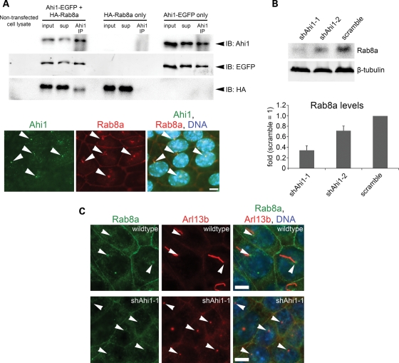
Figure 5.
Ahi1 is involved in ciliogenesis through its interactions with the small GTPase, Rab8a. (A, top) Co-immunoprecipitation of Ahi1-EGFP with HA-Rab8a in lysates from HEK293 cells transfected with Ahi1-EGFP and HA-Rab8a, but not in cells transfected with HA-Rab8a alone or Ahi1-EGFP alone. Resin only controls had no labelling (data not shown). (A, bottom) Co-localization of both Ahi1 (green) and Rab8a (red) at the basal body in wild-type cells (denoted by white arrowheads). (B) The protein level of endogenous Rab8a from Ahi1-knockdown (shAhi1-1 and -2) and from scramble shRNAi control cells was analysed by western blotting (top) and graphically displayed (bottom). The level of β-tubulin represents the loading control. The error bars represent the standard error of the mean. (C) Wild-type (top panel) and Ahi1-knockdown IMCD3 cells (lower panel) were stained with antibodies against Rab8a (green) and Arl13b (red; marker for the basal body and the primary cilium). Right panel represents the merged images of Rab8a, Arl13b and DNA (blue) staining. White arrowheads point to the basal body. With this Rab8a antibody, localization is only found at the basal body and does not show any apparent localization to the cilium. Scale bars =5 µm in (A and C).

