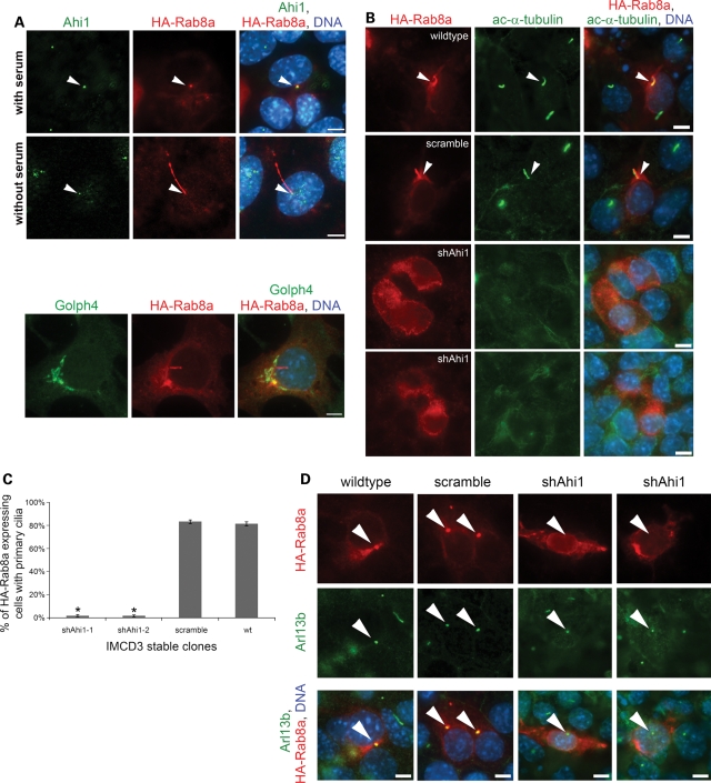
Figure 6.
Overexpression of HA-Rab8a in Ahi1-knockdown cells results in an inability to rescue ciliogenesis and an inability for HA-Rab8a to localize to the basal body of the primary cilium. (A) Wild-type IMCD3 cells, cultured under non-cilia forming conditions (top) or under serum-deprived conditions so as to promote cilium formation (bottom), and transfected with an HA-Rab8a expression plasmid were immunostained for Ahi1 (green) and for HA (red). Right panel represents the merged images of Ahi1, HA-Rab8a and DNA (blue) staining. White arrowheads point to the basal body. The bottom panel represents HA-Rab8a localization to the Golgi complex (using the Golgi marker Golph4 (green)). (B) Wild-type, scramble and Ahi1-knockdown cells transfected with HA-Rab8a were immunostained with acetylated α-tubulin (green) for primary cilia and HA for Rab8a (red) distribution. Arrowheads indicate the co-localization of HA-Rab8a and acetylated α-tubulin staining. Right panel represents the merged images of HA-Rab8a, acetylated α-tubulin and DNA (blue) staining. (C) Quantification of cilium formation in HA-Rab8a expressing wild-type, scramble and Ahi1-knockdown cells. Cells were co-stained with HA and Arl13b. Fifty HA-positive cells were counted from each slide and the number of primary cilium was determined. Data were collected from three independent experiments. The error bars represent the standard error of the mean. Asterisks indicate significance from scramble and wild-type cells (P < 0.0001) using Chi-square analyses. (D) Wild-type and scramble cells [grown to sub-confluence (resulting in few cilia); left two panels] and Ahi1-knockdown cells (grown under cilia optimized conditions; right two panels) transfected with HA-Rab8a were stained with antibodies against HA (red, for HA-Rab8a) and Arl13b (green, for the basal body). Arrowheads point to Arl13b labelled basal bodies. The lower panel represents the merged images of HA-Rab8a, Arl13b and DNA (blue) staining. This experiment was designed to capture wild-type and scramble cells at a time when few cells had cilia, so as to easily observe the association of HA-Rab8a in the basal body (before the formation of cilia where HA-Rab8a would enter the cilium making it difficult to differentiate basal body from the primary cilium). Scale bars =5 µm in (A, B and D).

