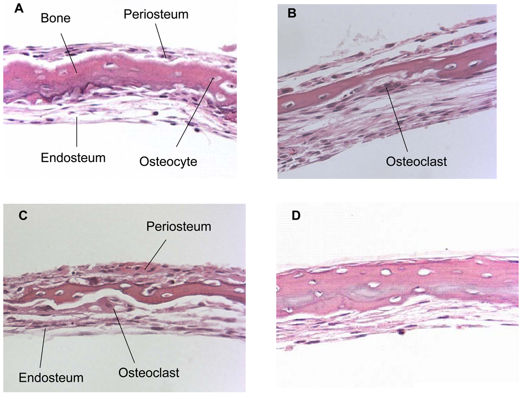
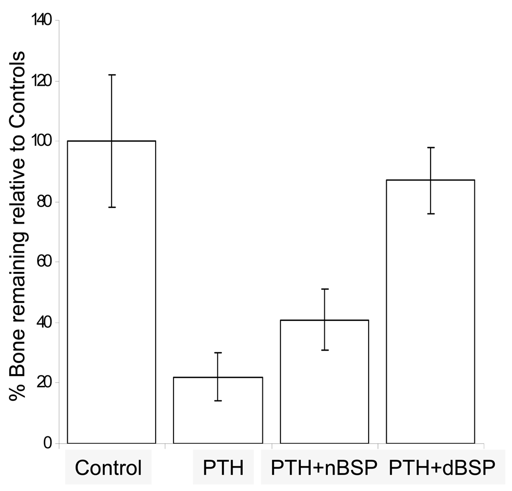
Figure 5. H&E stained paraffin sections of calvarial bones cultured for 7 days stimulated by PTH to resorb in the absence and presence of BSP forms and quantitative histomorphometric analysis.
Panel 1; A: control; B: in the presence of 10 nM PTH; C: in the presence of 10 nM PTH + 5 µg/ml of native BSP; D: in the presence of 10 nM PTH + 5 µg/ml of dephosphorylated BSP. Note the multinucleated resorbing osteoclast in B and C on the endosteal side. All the sections are at 25x magnification. Panel 2; Histomorphometric data for each of the groups represented by A–D expressed as % amount of bone remaining relative to the controls. Four different cultured calvaria for each group (a)–(d) were used with two sections per calvaria for computing the overall histomorphometric data. (a) the PTH alone treated cultures lost 77 ± 8 % of their mineralized bone matrix, (b) the PTH + native BSP cultures lost 58 ± 5 % of their mineralized bone matrix, and (c) the PTH + dephosphorylated BSP showed almost no loss of mineralized bone matrix, 11 ± 10 %.


