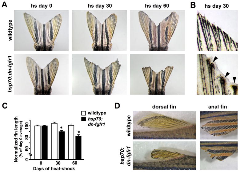
Fig. 1. Inhibition of Fgf signaling causes progressive tissue loss from zebrafish fins.
(A) Images of wildtype and hsp70:dn-fgfr1 fins at day 0, day 30, and day 60 of heat-shock. Wildtype fins were unaffected by daily heat-shocks, while transgenic fins showed progressive loss of distal fin tissue. Fins shown are representative, and not from the same animal at each timepoint. (B) High magnification images of distal fin structures after 30 days of heat shock. Many hsp70:dn-fgfr1 rays exhibited severe tissue loss, which was often accompanied by an excess of epidermal tissue (arrowheads). (C) Quantification of fin loss by measurement of centrally located rays (see Materials and methods). hsp70:dn-fgfr1 animals displayed significant reductions in fin length following both 30 and 60 days of heat-shock, whereas wildtype clutchmates showed no changes (mean ± SEM; *Student’s t-test, p ≪ 0.001 at days 30 and 60). (D) Dorsal and anal fins of hsp70:dn-fgfr1 zebrafish showed fin atrophy after 60 days of Fgfr blockade. The fins of wildtype clutchmates retained their length and morphology after 60 days of similar heat treatments.

