
Figure 4.
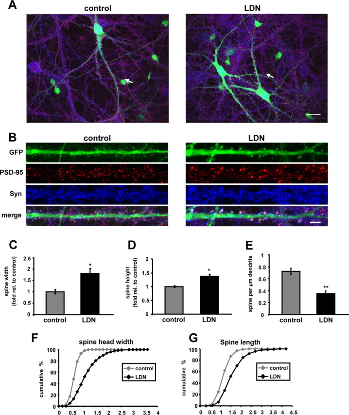
Inhibition of UCH-L1 activity alters spine size and density. A, B, Cultured neurons were treated with LDN for 24 h. EGFP Sindbis virions were added directly to culture media after 10–12 h of LDN treatment, and protein expression was allowed to continue for 12–14 h. At the end of LDN treatments, neurons were fixed, permeabilized, and immunolabeled with anti-PSD-95 and anti-Synapsin I antibodies. The straightened dendrites in B correspond to the regions indicated by arrows in the whole-cell images in A. C–G, Quantification of the number, widths, and lengths of GFP-filled spines in control and LDN-treated neurons is shown. C, D, Measurements for spine head widths and spine lengths were normalized to those of control values. F, G, Cumulative frequency plots showing distribution of spine head width (in micrometers) and spine length (in micrometers) in control and LDN-treated neurons are shown. E, Quantification of spine density is represented as the number of spines per 1 μm dendrite length. The quantified data were obtained from three independent experiments, in which >20 dendrites and >900 spines were analyzed per condition. Mean values ± SEM are shown. *p <0.05, **p < 0.01, unpaired Student's t test.