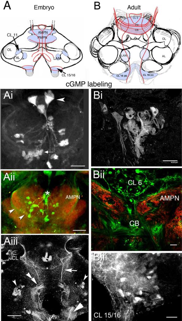
Fig. 4.
Localization of cyclic 3,5 guanosine monophosphate (cGMP) in the embryonic (E85%) and adult brain when NO is up-regulated with SNP and IBMX. cGMP antibody is revealed in green in the double-labeled images. The colabel, synapsin (red), delineates brain structures. A,B: Schematic diagrams of embryonic (A) and adult (B) stages indicating neuropil regions and cell clusters that label for NOS (light blue). The red lines in A and B represent longitudinal fibers and fibers that cross the brain at the level of the PB and CB. Some fibers lie in the protocerebral tracts. Ai: By E85%, three components of the frontal eye (arrowhead) label. A second cluster of smaller photoreceptor cells also label (arrow). Aii: cGMP antibody photoreceptor cells and AMPN (punctate labeling, arrowheads). The asterisk marks the base of the naupliar eye. Aiii: Medial tracts (arrow) in the embryo (in the same location as NOS-labeled fibers at this same stage), as well as a commissure that projects to the eyestalks (asterisk), label for cGMP. Also stained are cells in CL 11 (small bilateral arrowheads) and 15/16 (large arrowhead). The double arrowhead marks the deutocerebral fan-shaped collection of fibers whose origin has not been identified. Bi: In the adult brain, photoreceptor cells label in the protocerebrum; the three large components of the frontal eye do not. Bii: In fibers in the CB, AMPN and CL 6 label. Biii: Labeled cells in CL 15/16 have fibers that extend into the connectives. For abbreviations, see list. Scale bars = 80 μm in Ai,Aiii, 50 μm in Aii, 40 μm in Bi, 50 μm in Bii,Biii.

