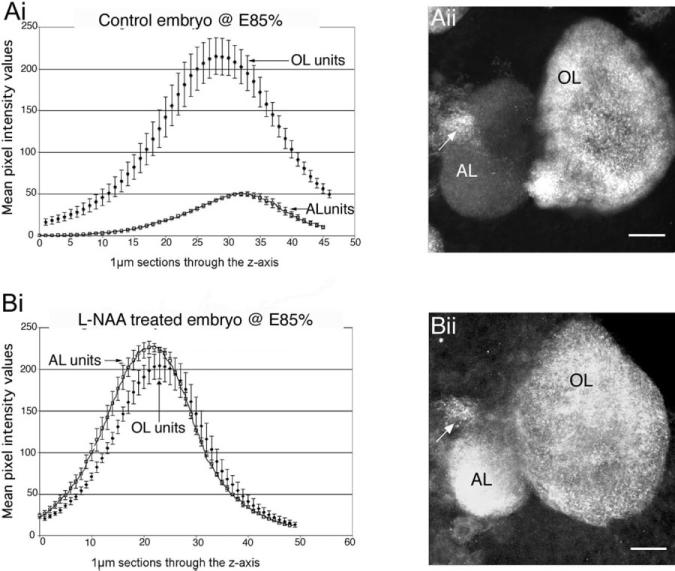
Fig. 8.
Aii: Synapsin labeling is not present in the accessory lobes in control embryos (see also Fig. 7A). Bii: However, after L-NAA treatment (down-regulation of NO) of E85% embryos, there is intense synapsin labeling in the AL. Ai, Bi: Graphs show the relative intensity levels of synapsin labeling in the OL and AL in embryonic brains at E85%; Control, n = 10 (Ai), NOS-inhibited, n = 10 (BI). Bi: Semiquantitative measurements demonstrate that the intensity of synapsin staining has indeed increased following L-NAA treatment and is comparable to the intensity of labeling in the OL. Each graph represents the average mean intensity values of the two lobes ± the SD through the z-axis of a stacked confocal image. Designated regions of interest in the confocal stacked images of the control (Ai) and NOS-inhibited (Bi) brains were analyzed with Leica software to produce the data for the graphs. The arrows point to the OGTN, which labels in both conditions. For abbreviations, see list.

