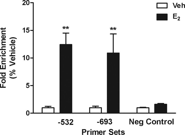
Figure 6.
Results of ChIP using ERα antibody and DNA isolated from N42 cells. Cells were treated with ethanol vehicle (Veh) or E2 for 2 h before they were harvested for ChIP assays. Samples were assayed using primers flanking the −532 ERE and the −693 ERE or a site with no ERE consensus sequences (for primer sequences, see Materials and Methods). Each data point represents the mean ± SEM of three independent samples, each analyzed in duplicate. **Significantly different from corresponding vehicle control; p < 0.01. Neg, Negative.

