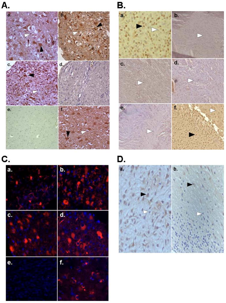
Figure 2. Detection of HHV-6A/B gp116/64/54 Late Antigen by Immunohistochemistry and Immunofluorescence microscopy in Pediatric Brain Tumors and Control Brain.

A. Immunohistochemistry using HHV-6A/B gp116/64/54 late antigen antibody of PCR positive pediatric tissue microarray gliomas (a-d) at 20× magnification reveals both positive (black arrows) and negative (white arrows) containing regions. Staining is preferentially observed in lower grade gliomas (a-c) and HHV-6 encephalitis positive control (f) compared to high grade glioma (d) or non tumor control tissue (e). B. HHV-6A/B gp116/64/54 late antigen staining is observed in an oligodendroglioma (a) but not in other non glial tumor subtypes meningioma (b,c), medulloblastoma (d), or germinoma (e) compared to positive control HHV-6 encephalitis (f). C. Immunofluorescence microscopy using HHV-6 A/B gp116/64/54 late antigen further reveals cytosolic signal in PCR positive gliomas (a-d) but not age-matched control brain (e) compared to HHV-6 encephalitis positive control (f). D. HHV-6A/B gp116/64/54 IHC of a pediatric glioma containing tumor (a) and adjacent non tumor (b) areas reveals increased signal in the tumor containing region.
