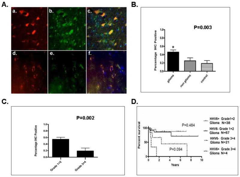
Figure 3. Detection of HHV-6A/B gp116/64/54 Late Antigen in Cells of Glial Origin and Correlation between Tumor Type, Grade, and Progression Free Survival.

A. Immunofluorescence microscopy of pediatric glioma (a-c) and HHV-6 control encephalitis (d-f) paraffin embedded samples dual stained with HHV-6A/B gp116/64/54 late antigen(a,d) and glial fibrillary acidic protein (b,e) reveals colocalization (c,f) indicative of HHV-6 antigen in cells of glial origin. B. Statistical analysis of gp116/64/54 IHC results shows predilection of immunopositivity in glial tumors compared to non glial tumors (P<0.003). C. HHV-6A/B late antigen gp116/64/54 is preferentially detected in lower grade (1-2) gliomas compared to higher grade gliomas (3-4) (P<0.002). D. Kaplan Meier progression free survival analysis stratified by HHV-6A/B gp116/64/54 IHC status and tumor grade demonstrates no significance among IHC positive or negative lower grade (P=0.484) or higher grade (P=0.094) gliomas.
