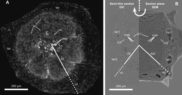
Figure 1.
Morphology of a detached T. wilhelma bud (stage 4). Comparison between (A) a virtual section from an SR-μCT data set and (B) 'combined scanning electron histology' data consisting of a semi-thin section imaged using DIC-microscopy and SEM of the corresponding surface after sectioning (cc - choanocyte chamber, cd - choanoderm, co - cortex, dcd - developing choanoderm, lac - lacunae, msb - megasclere bundle).

