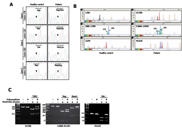
Figure 2.
Mutations in PRNP were detected from probable CJD patients. Electropherogram of the DNA sequences (A), SNaPshot assay (B), and digested PCR fragments with restriction enzymes as indicated (C) revealed the mutations at D178N (GAC→AAC), E200K (GAG→AAG)-129MV (ATG→GTG), and M232R (ATG→AGG). (A) Arrow heads indicate the mutated nucleotide peaks. (B) The x and y axes represented the size of bases and the fluorescence intensities, respectively. The color of each peak indicates the identity of the ddNTPs in the primer extension reaction, emitting specific fluorescences according to ddNTP; Green/A, Black/C, Blue/G, and Red/T. The orange peaks represented the internal size standard. The arrows labeled "129" or "200" indicate SNaPshot products resulting from the extensions of the S129 and S200 primers, respectively.

