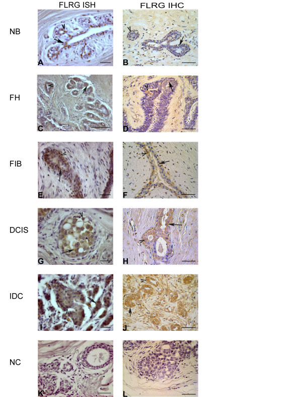
Figure 3.
FLRG representative microscopic views of: mRNA (A,C,E,G,I) and protein (B,D,F,H,J) expression detected by in situ hybridization (ISH); and immunohistochemistry (IHC), respectively, in normal breast (NB), florid hyperplasia (FH), fibroadenoma (FIB), ductal carcinoma in situ (DCIS) and infiltrative ductal carcinoma (IDC). FLRG staining was detectable in the epithelial cytoplasm (arrow) and nucleus (arrowhead) in all the cases analyzed, with some weak staining detected in the stromal cells. K and L: negative controls for ISH and IHC respectively. Bar 50 μm.

