
Fig. 3.
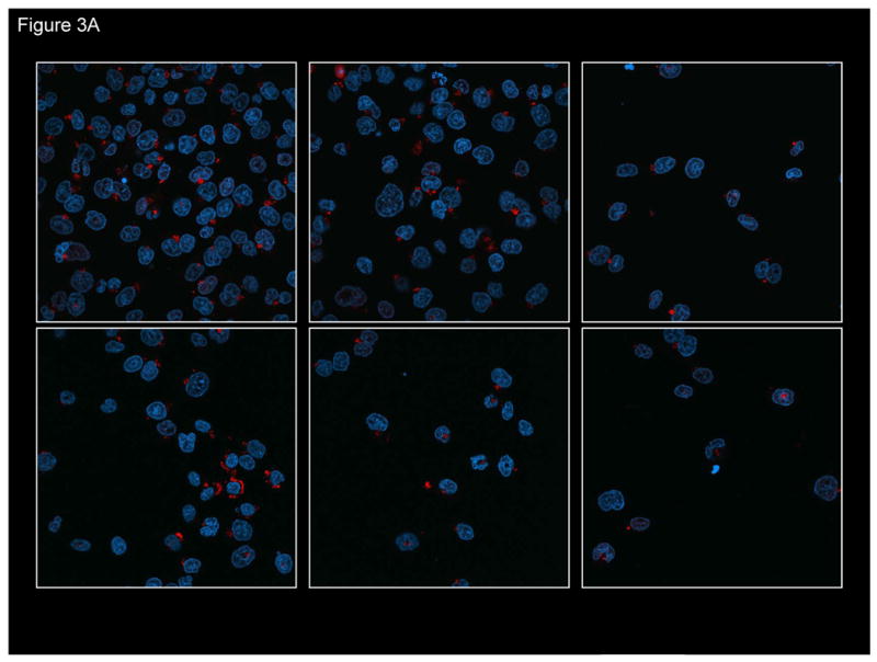
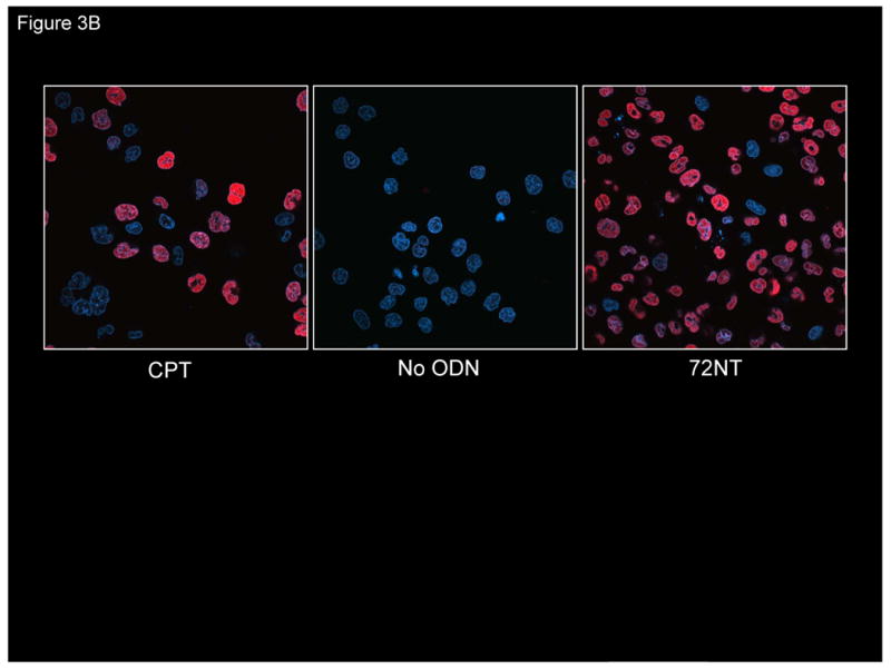
(A) Confocal image of fluorescently-tagged oligonucleotide in and around the cell nucleus. HCT116-19 cells were electroporated with 8 μM of Cy3-labeled ODN (red) and allowed to recover for 24 hours. DAPI was used to label nucleus (blue). (B) Presence of ssODN stimulates H2AX activation. Confocal image of pH2AX in HCT116-19 cells in the presence of 8 μM 72NT. Camptothecin (CPT) (300 nM) treatment serves as positive control.