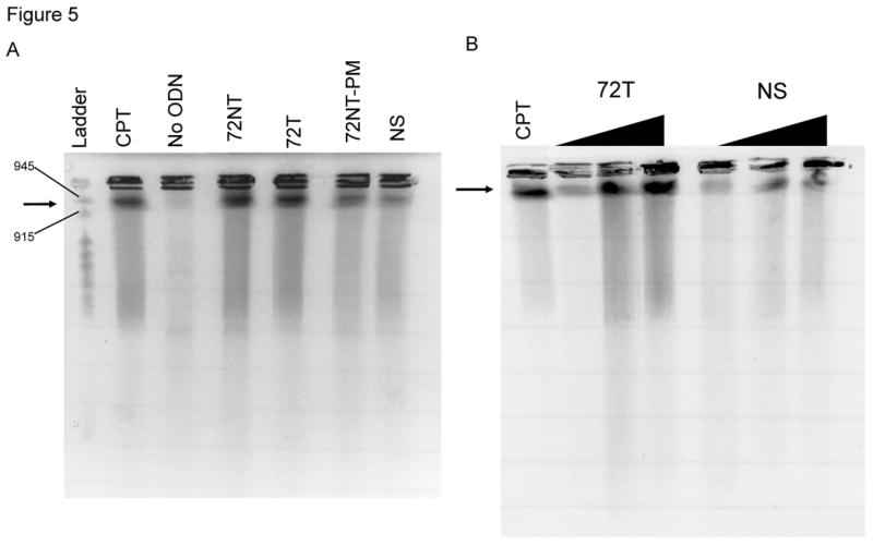
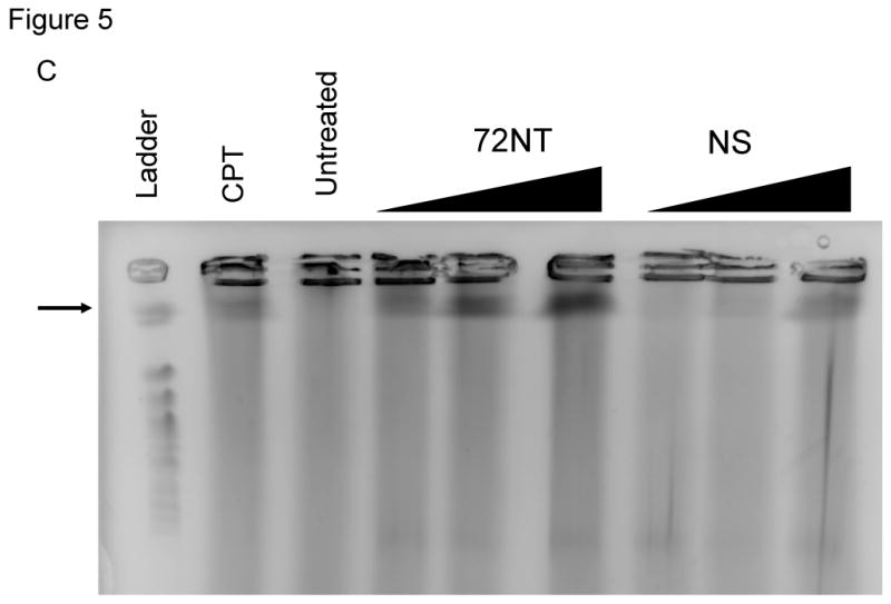
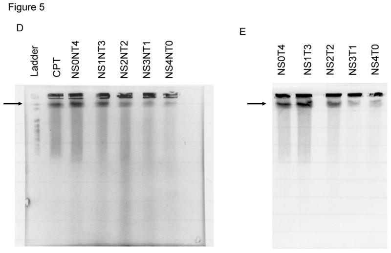
Fig. 5.


(A) Pulse-field gel electrophoresis (PFGE) of HCT116-19 cells 24 hours after being electroporated with 4 μM of respective ODN after 24 hour pre-treatment with 2 μM aphidicolin. A non-specific (NS) ODN does not induce double-strand breaks, whereas ODNs specific to either the NT or T strand do. (B) PGFE of HCT116-19 cells, 24 hours after electroporation, shows DSB formation is dose-dependent. 72NT and NS ODNs were present at 2, 4, and 8 μM. (C) PFGE of HCT116-19 cells, 24 hours after electroporation, with 4 μM total ODN (72NT and non-specific) in specified molar ratios. (D) Pulse-field gel of HCT116-19 cells, 24 hours after electroporation, with 4 μM total ODN (72T and non-specific) in specified molar ratios. Increasing the molar ratio of NS ODN relative to either the 72NT or 72T ODN decreases the amount of DSBs.
