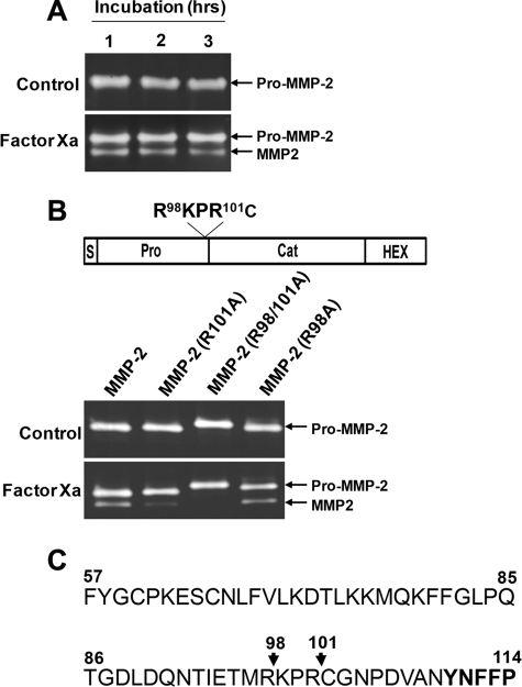
FIGURE 3.
Factor Xa cleavage sites in the MMP-2 propeptide. A, direct cleavage of the propeptide by factor Xa. Purified pro-MMP-2 (100 ng/ml) from transfected COS-1 cells was incubated with 50 nm factor Xa for the indicated times, and zymographic analysis was performed. The arrows indicate pro-MMP-2 and processed MMP-2. B, zymograms of conditioned medium from COS-1 cells expressing pro-MMP-2, pro-MMP-2 R101A, pro-MMP-2 R98A/R101A, or pro-MMP-2 R98A treated with or without 50 nm factor Xa for 16 h. Note that the pro-MMP-2 R98A/R101A mutant migrates slower on the gel, possibly due to the replacement of two positively charged Arg residues with the uncharged Ala. The drawing at the top shows the mutation sites. C, partial sequence of the propeptide and start of the catalytic domain (boldface type). The arrows indicate factor Xa cleavage sites (n = 3).

